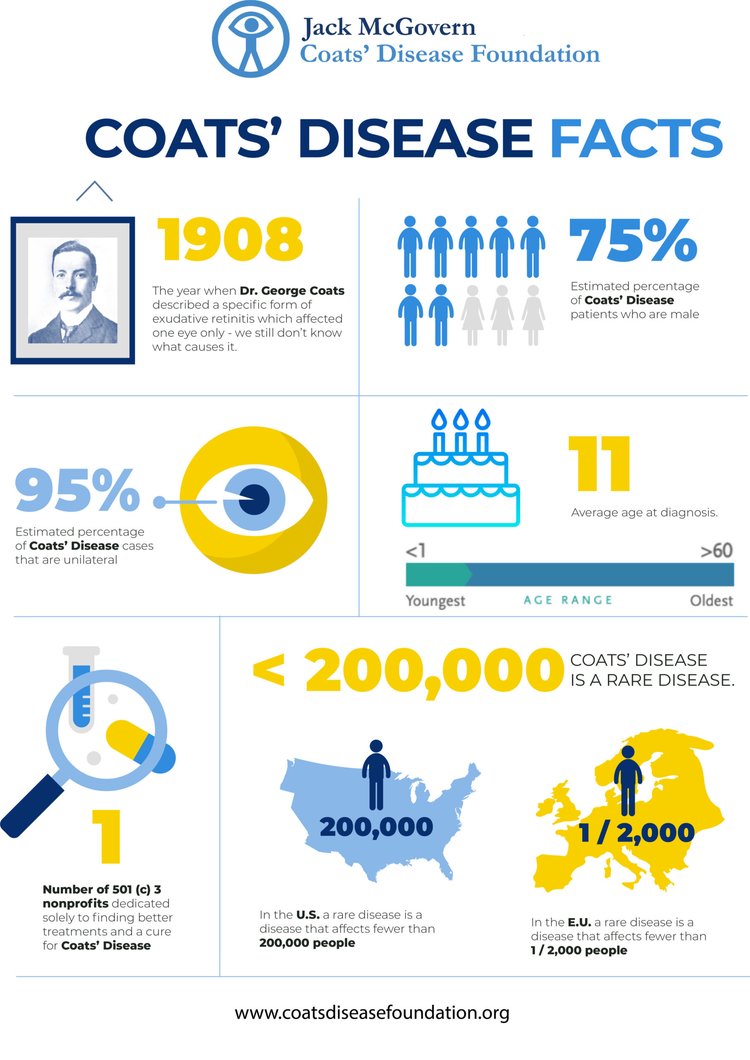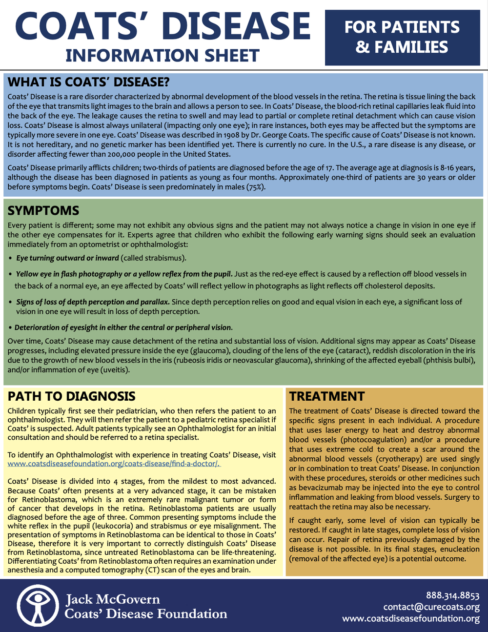
Coats’ Disease, or exudative retinitis or retinal telangiectasis, sometimes spelled Coates Disease, was first identified by Scottish ophthalmologist George Coats in 1908. It is a very rare condition where there is abnormal development in the blood vessels behind the retina. The blood rich retinal capillaries break open, leaking the serum portion of the blood into the back of eye. The leakage causes the retina to swell, and can cause partial or complete detachment of the retina.
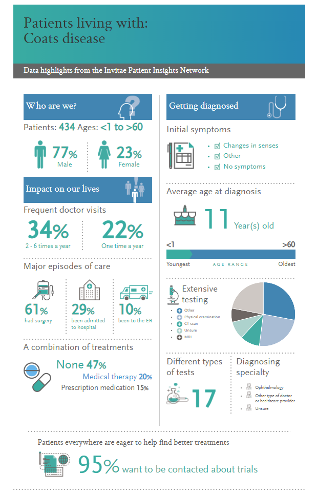
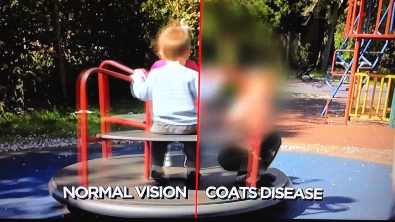
Coats’ Disease is typically diagnosed in childhood. Because of lack of disease awareness, even in the medical community, the vision can become very poor in the affected eye prior to diagnosis. Infants and young children may develop misalignment of their eyes (strabismus) or a “white reflex” from the pupil (leukocoria meaning "white pupil") in the affected eye (as opposed to the normal “red reflex” one sees when light is shined into the eye or from a camera flash). https://www.aapos.org/glossary/leukocoria
Children typically first see their pediatrician who then refers the patient to an ophthalmologist. They will then refer the patient to a pediatric retina specialist if Coats’ is suspected.
Coats’ Disease is divided into 4 stages from the mildest to most advanced. Even with stage 2 disease, half of eyes become legally blind despite treatment. Currently, treatment for Coats’ disease involves laser to the retina and medicine injections into the eye to repair the retinal detachment and scarring. There is no cure for Coats’ and repair of retina previously damaged by the disease is not possible.
Because Coats’ often presents at a very advanced stage, it can be mistaken for cancer in the eye. Differentiating Coats’ from a childhood eye cancer known as retinoblastoma often requires an examination under anesthesia and a computed tomography (CT) scan of the eyes and brain. The process of reaching the diagnosis of Coats’ can be very stressful to the patient and their family. In its advanced form, severe blindness is very common. Even when discovered later in life, central vision loss is common.
Coats’ Disease is seen predominantly in males (69%) and symptoms generally appear in children aged six to eight but can develop as young as four months. It progresses gradually and affects central vision. It is almost always unilateral (affects only one eye). If caught early, some level of vision can typically be restored. If caught in late stages, complete loss of vision can occur. In its final stages, enucleation (removal of the affected eye) is a potential outcome.